Personalized Prosthesis Using 3D Printing for Complex Hip Revision
This page explains how a personalized prosthesis using 3D printing is built when bone stock is gone and off-the-shelf cup and stem sizes no longer fit. It covers the data chain, the print parameters that matter, and where machining takes over. Engineers and sourcing teams can use it to judge whether a case is a print job or a standard part job.
In this article
- 1
- 2
- 3
- 4
- 5
When a Personalized Prosthesis Using 3D Printing Beats a Catalog Implant
A primary hip replacement works because the surgeon can pick a cup and stem from a size chart. The bone is intact enough to hold them. A revision after one or two failed implants is a different problem. The acetabulum may have lost its medial wall, the columns may be cracked, and the femur may be missing the metaphyseal bone that a standard stem needs for fixation.
When the defect gets large, no catalog part matches the remaining bone. The surgeon either rebuilds the defect with bone graft and cages, or removes the guesswork and fits an implant to the anatomy that is actually left. That second route is where a personalized prosthesis using 3D printing earns its place.
The decision is not about novelty. It is about contact area and load path. A printed cup with a patient-matched flange can sit on the ilium and ischium where bone still exists. A standard hemispherical cup needs a rim to press into. If the rim is gone, the cup has nothing to grip.
Size is the second gate. Revision stems are offered in a limited range of lengths and diameters. When the canal is wider than the largest available stem, or the offset needed is outside the catalog, the fit becomes a compromise. A printed body can follow the canal and place the joint center where the soft tissues want it.
- 1Large uncontained defectsBone loss beyond what a hemispherical cup can bridge.
- 2Pelvic discontinuityColumns cracked or separated; the implant must span the gap.
- 3Canal wider than the largest stemStandard stems cannot achieve a scratch fit.
- 4Joint center off by more than 10 mmLeg length and abductor tension need a patient-matched offset.
From CT Scan to Printable Geometry
Everything starts with imaging. A thin-slice CT scan, usually 0.5–1 mm slices, is segmented to separate bone from soft tissue and from metal artifact left by the old implant. Metal scatter is the first real obstacle. Without artifact reduction, the bone surface near the failed cup looks like a cloud, and any surface built on it will be wrong.
The segmented bone becomes a surface model. The engineer then mirrors the healthy side to estimate where the original joint center sat, and overlays that target on the damaged side. This step sets the offset, the version, and the leg length. Get it wrong here and the whole implant is wrong, no matter how well it is printed.
Next comes the implant body itself. It is not a solid block. Solid titanium is stiff, and a stiff implant next to relatively flexible bone takes almost all the load. That leads to stress shielding and bone resorption around the implant. The fix is a lattice or porous region at the bone interface, with a solid core only where screws and the joint bearing need material.
Finally the design is checked against the surgical plan. Screw corridors must pass through bone, not through the bladder or the great vessels. Flanges must not impinge on the sciatic nerve. This review is a joint effort between the surgeon and the manufacturing engineer, and it usually runs two or three rounds before the file is locked.
- 1Slice thickness0.5–1 mm; thicker slices blur the acetabular rim.
- 2Metal artifact reductionRequired when the old implant is still in place.
- 3MirroringUses the contralateral side to restore the joint center.
- 4Screw corridor checkEvery hole must land in bone with a safe margin.
Printing, Heat Treatment, and Machining the Interfaces
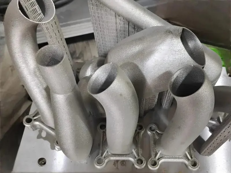
The build runs on a laser powder bed fusion machine using Ti-6Al-4V (TC4) powder. Layer thickness sits between 30 and 60 μm. Thinner layers give a smoother surface and better resolution on fine lattice struts, but they slow the build. For a cup-sized part, a 30 μm layer is common; for a large revision cage, 50–60 μm keeps the build time practical.
Laser power, scan speed, and hatch spacing are tied together. The goal is full density with no lack-of-fusion pores. Pores at the surface become crack initiation sites under cyclic load, and a hip implant sees millions of cycles. Melt pool monitoring during the build flags any layer where the signal drifts outside the qualified window.
After the build, the part is stress relieved and then hot isostatically pressed. HIP closes internal porosity and improves fatigue life. The part is then cut from the build plate and the support structures are removed. Support removal on a lattice region is delicate work. Cutting too deep removes struts; leaving too much leaves rough material that cannot be cleaned.
Printed surfaces are not final surfaces. The bearing taper, the screw holes, and any mating face are machined. On our 5-axis centers we hold ±0.005 mm (±0.0002 in) on those features and finish them to Ra 0.8–1.6 μm. The porous bone-contact region is left as-printed, because that roughness is what bone grows into. Blasting or polishing it would destroy the function.
Cleaning follows. Loose powder trapped in lattice channels has to come out. The part goes through ultrasonic baths and, where the geometry demands it, pressure washing from multiple angles. Powder left inside a lattice can migrate after implantation and cause third-body wear at the bearing. This is a documented step, not a rinse.
- 1Layer thickness30–60 μm depending on feature size and build time.
- 2Build atmosphereArgon, with oxygen kept low to avoid oxidation.
- 3HIPCloses porosity and lifts fatigue strength.
- 4Machined featuresTaper and screw holes held to ±0.005 mm.
Material Choice and the Tests That Close the Loop
Titanium is the default for load-bearing revision implants. Ti-6Al-4V (TC4) has the fatigue strength and the elastic modulus that make a porous lattice viable. Commercially pure TA1 and TA2 are softer and are used for non-load-bearing plates and covers rather than for a cup that carries body weight every step.
The powder itself is a controlled input. Particle size distribution, chemistry, and oxygen content are checked on receipt, and powder is reused only within a validated number of cycles. Oxygen pickup during reuse raises brittleness. A supplier that tracks reuse without a limit is a risk, no matter how good the printer is.
Testing runs at three levels. Powder and build coupons verify the process. Coupons are printed with the part and tested for density, chemistry, and tensile properties. Then the finished part is checked dimensionally against the design model, usually on a coordinate measuring machine for the machined features and by CT for internal lattice geometry.
Traceability ties it together. The build file, the machine, the powder lot, the heat treatment batch, and the inspection records all attach to one device history record. For a medical device this is not optional. If a question comes up two years later, the answer has to be findable.
One limit worth stating plainly. A printed implant solves fit and fixation. It does not solve infection. If there is active infection at the site, the revision is usually staged, with a spacer first and the definitive implant later. Printing a custom part into an infected bed is the wrong use of the technology.
- 1Ti-6Al-4V (TC4)Load-bearing cups, cages, and stems.
- 2TA1 / TA2Non-load-bearing covers and plates.
- 3Powder reuse limitValidated cycle count, oxygen monitored.
- 4Device history recordBuild, lot, heat treat, and inspection linked.
Catalog Implant or Personalized Prosthesis: Which Case Fits Which
A quick screen for the revision planning meeting
| Case feature | Catalog implant | Personalized prosthesis using 3D printing |
|---|---|---|
| Acetabular defect | Contained, rim intact | Uncontained, rim absent |
| Bone contact | Hemispherical press fit | Flange on remaining columns |
| Canal diameter | Within stem size chart | Wider than largest stem |
| Joint center offset | Restored within 10 mm | Needs patient-matched offset |
| Lead time | Days, off the shelf | Weeks, design and build |
| Cost per part | Lower, catalog pricing | Higher, single-unit build |
| Best use | First revision, good bone | Complex revision, poor bone |
The Call We Would Make
If the defect is contained and a catalog cup still gets rim contact, use the catalog part: it ships in days and costs less. Go to a personalized prosthesis using 3D printing when the bone cannot hold a hemispherical cup, the canal is wider than the largest stem, or the joint center cannot be restored with standard offsets. Fit and fixation are the reasons to print, not convenience.
Questions We Get From Revision Teams
How long does the whole process take, from CT to finished part?
Design and review usually take the longest, because the surgeon and the engineer go back and forth on screw corridors and flange placement. The build itself is a matter of days, and machining the taper and screw holes adds a short step after that.
We quote and return a free DFM analysis within 12 hours of receiving the files, and production can start within 24 hours once the design is locked. The schedule driver is design approval, not machine time.
Can you print the bearing surface too?
We print the metal body, including the porous bone-contact region. The articulating bearing is a separate concern. A metal-on-polyethylene or ceramic bearing pair is normally used, and the metal taper that locks the bearing in place is machined, not printed.
That machined taper is held to ±0.005 mm and finished to Ra 0.8–1.6 μm. Printing it would leave surface texture and porosity where a precise fit is required.
What porosity level is right for bone ingrowth?
Porous regions are typically designed with 60–80% porosity and pore sizes in the 300–800 μm range. Smaller pores restrict vascularization; larger ones reduce the strength of the lattice and its contact with bone.
The exact number is a design decision tied to the load the region carries. A cup dome that takes compressive load is not built the same as a non-load-bearing cover plate.
Does the part need machining at all if it is printed near net shape?
Yes, on the functional interfaces. Printed surfaces have a roughness that comes from the powder bed, and dimensional tolerance on an as-printed face is looser than what a taper or a screw hole needs.
We machine those features on 5-axis centers. The porous bone-contact region stays as-printed on purpose, because that texture is the point.
How is confidentiality handled for patient imaging data?
Uploads are secure and confidential, and we sign an NDA on request. Imaging data and design files are kept inside the project record and are not shared outside the people working on the case.
Our information security management system is certified to ISO 27001:2022, and our medical device quality system is certified to ISO 13485:2016.
Can you run a single unit, or is there a minimum order?
There is no minimum order quantity. A revision case is a single unit, and we run it as one. The same lines also handle runs from one prototype up to 10,000+ parts for instrument sets and other hardware.
Every part gets 100% inspection before shipment, with raw material checks, in-process monitoring, and a final dimensional report on request.
Send Us the Case Files
Upload the CT data and the surgical plan. You get a quotation and a free DFM analysis within 12 hours, and an engineer reviews the design with you before anything is built.
12-hour quote100% inspectionISO 13485:2016NDA on request
More From the Shop Floor
We publish setup notes, tooling trials and inspection data from the factory floor.